THURSDAY, June 12, 2014 (HealthDay News) — A simple and quick MRI technique might aid in early detection of Parkinson’s disease, British researchers report.
The new MRI approach can detect with 85 percent accuracy people who have early stage Parkinson’s disease, according to findings published online June 11 in the journal Neurology.
That’s important because the early symptoms of Parkinson’s are subtle, which makes an accurate early stage diagnosis very difficult, the researchers said.
This new test, which focuses on scans of a part of the brain known as the basal ganglia, could improve the lives of countless patients with Parkinson’s, said senior author Clare Mackay, a senior research fellow with the Oxford Parkinson’s Disease Center at Oxford University.
“By the time symptoms of Parkinson’s are obvious, a lot of damage has already taken place in the brain,” Mackay said. “To reduce the impact of Parkinson’s we need to find new ways to protect the brain before significant damage is done, so we must have good tools to identify people as early as possible in the course of the disease.”
Parkinson’s disease is a progressive disorder of the nervous system that occurs when the body fails to produce enough of a brain chemical called dopamine, according to the National Parkinson Foundation. Early symptoms include trembling, stiffness and poor coordination, but proceed to the point where patients may have trouble walking, talking, chewing, swallowing or speaking.
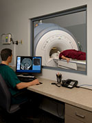
Conventional MRI can’t detect early signs of Parkinson’s, so the researchers turned to an MRI technique called resting-state functional MRI.
Patients are required to stay still in the scanner while doctors examine the strength of the brain networks linked to the basal ganglia, the deep brain region associated with Parkinson’s disease.
A different type of brain scan, PET, already has shown promise as an early detection tool for Parkinson’s, but the test is very expensive and involves giving people a dose of radiation, Mackay said.
“MRI is relatively inexpensive and widely available,” she said, adding that the test’s lack of radiation allows for safer screening and repeat scanning.
The team developed the technique by comparing 19 people with early stage Parkinson’s disease while not on medication with 19 healthy people, matched for age and gender. The investigators found that the Parkinson’s patients had much lower connectivity in the basal ganglia.
The researchers then applied their MRI test to a second group of 13 early stage Parkinson’s patients, correctly identifying 11 out of the 13 patients — an accuracy score of 85 percent.
According to Mackay, “85 percent is promising, and we’re working on ways to improve this further.”
The Oxford Parkinson’s Disease Center treats more than 1,000 patients, which provides ample opportunity for researchers to collect new scans, she said.
“We’re also collecting scans from people who are at increased risk of Parkinson’s, and will follow them up to find out whether our method could reliably predict who would develop the disease,” Mackay noted.
That said, Mackay added that “brain scanning would never be used to diagnose Parkinson’s in isolation. It is likely that the best sensitivity will come from combining brain scanning with other measurements.”
The medical director of the National Parkinson Foundation, Dr. Michael Okun, hailed the results of the new MRI test.
“It is very encouraging to see scientists attempting to validate simpler and more widely available techniques for detecting early Parkinson’s disease,” Okun said. “These sorts of approaches, if validated by larger better-powered studies, could be instrumental when applied to trials of potentially disease-modifying therapies.”
More information
For more about Parkinson’s disease, visit the U.S. National Library of Medicine.
Copyright © 2026 HealthDay. All rights reserved.

