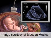
THURSDAY, Feb. 21 (HealthDay News) — The human heart is not fully formed until much later in pregnancy than previously thought, a new study suggests.
British researchers analyzed scans of the hearts of healthy fetuses in the womb and found that the heart has four clearly defined chambers in the eighth week of pregnancy, but does not have fully organized muscle tissue until the 20th week.
This is much later than expected, according to the study published Feb. 20 in the Journal of the Royal Society Interface Focus.
Previous studies of early heart development in humans have been largely based on other mammals — such as mice or pigs — as well as adult hearts and samples from dead people.
The data being collected by the University of Leeds-led team is being used to create a computerized simulation of fetal heart development. This will help improve understanding about normal heart development in the womb, and could lead to new ways to detect and treat some fetal heart problems early in pregnancy, according to the researchers.
Human fetuses have a regular heartbeat beginning at about 22 days of pregnancy, which is one reason why the researchers were surprised to find that there is little organization of human heart cells until 20 weeks of pregnancy.
“For a heart to be beating effectively, we thought you needed a smoothly changing orientation of the muscle cells through the walls of the heart chambers. Such an organization is seen in the hearts of all healthy adult mammals,” Dr. Eleftheria Pervolaraki, a visiting research fellow at the University of Leeds’ School of Biomedical Sciences, said in a university news release.
“Fetal hearts in other mammals such as pigs, which we have been using as models, show such an organization even early in gestation, with a smooth change in cell orientation going through the heart wall. But what we actually found is that such organization was not detectable in the human fetus before 20 weeks,” she explained.
More information
The Nemours Foundation offers a fetal development calendar.

