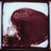
THURSDAY, Sept. 8 (HealthDay News) — New X-rays revealed the best and most accurate images of the brain shape of an early human ancestor, according to researchers.
The images of the fossil of an ancient species, known as MH 1 (Australopithecus sediba), shed new light on the evolution of the human brain, the study authors explained in the paper published Sept. 9 in the journal Science.
The well-preserved MH 1 fossil was discovered by Lee Berger from the University of the Witwatersrand in Johannesburg, South Africa, back in 2009. The cranium of the fossil was scanned at high resolution at the European Synchrotron Radiation Facility in France. Berger noted in a news release from the facility that the advanced features of the brain made it “possibly the best candidate ancestor” for the human genus.
The researchers pointed out that humans have a very large brain relative to their body size, about four times that of chimpanzees. Although the reconstructed volume of the MH 1 cranium was surprisingly small, they found its overall shape resembles humans more than chimpanzees.
The scientists also concluded that the X-ray images are consistent with a model of gradual brain reorganization in the front part of the brain.
The paper is included in a five-part series conducted by an international group of scientists about various parts of the Australopithecus sediba anatomy.
More information
The U.S. National Institute of Neurological Disorders and Stroke has more about the human brain.

